Organisms respond to changes in their internal and external environments (AQA A2 Biology) PART 6 of 9 TOPICS
|
Skeletal muscles are stimulated to contract by nerves and act as effectors:
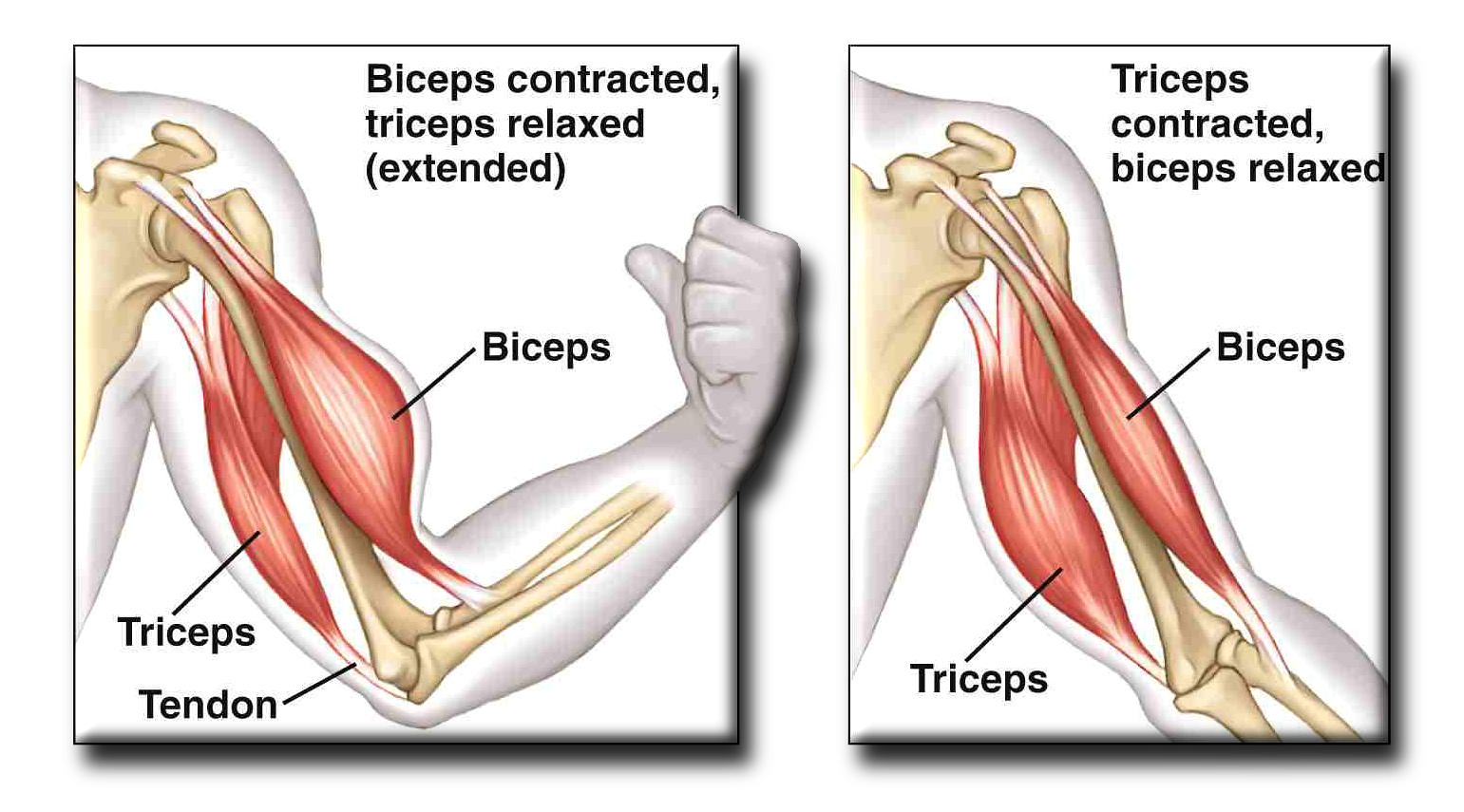
Muscles act as antagonistic pairs against an incompressible skeleton where if one muscle contracts (agonist) it pulls the bone and the other muscle relaxes (antagonist). An example is biceps and triceps in the arm. NB: Muscles do not push bones and only pull. Skeletal muscles are attached to bones by tendons. Ligaments are attached from one bone to the other.
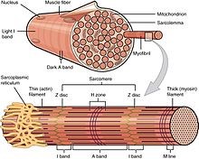
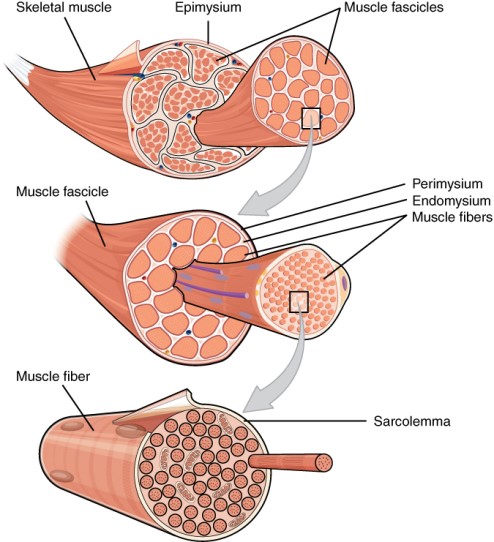
The gross structure is as follows: The muscle is made up of bundles which are packaged together. These bundles contain fibres/fascicles which are packaged in a connective tissue and have a distinct stripy look called striated tissue. These fibres/fascicles are known as muscle cells and have a membrane called sarcolemma, a cytoplasm called the sarcoplasm and organelles called myofibrils. These myofibrils are made up of 2 types of proteins: actin and myosin.
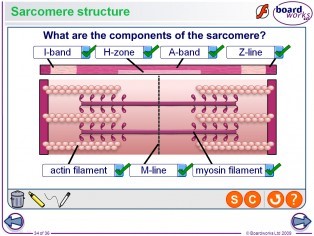
The microscopic structure should be known which is of the sarcomere. This is from one Z-line to another Z-line and is one contractile unit of many in a myofibril which are long and cylindrical in shape which shorten to create contraction. Between the two Z-lines in the middle is a line called the M-line. Adjacent to the Z-lines inside are I-bands (light bands), one on each side, which are lighter in colour as they only contain actin. The A-band (dark band) is from one I-band to the other and is darker in colour as it contains actin and myosin. H-zone is in the middle of the A-band and contains only myosin.
The sliding filament theory is as follows: At a neuromuscular junction on a fibre, an action potential is made and travels along the postsynaptic membrane and into the t-tubules which are indentations of the muscle cell. Sarcoplasmic reticulum, located at the t-tubules, release Ca2+ ions into the sarcoplasm when an action potential passes and is used to bind to tropomyosin which is wrapped around actin covering myosin-head binding sites. When it does bind to tropomyosin, it changes shape to expose the myosin-head binding sites so that the myosin heads can attach to form actinomyosin bridges. NB: The myosin heads actually bind to a protein called troponin on tropomyosin but this does not need to be known for AQA. The myosin heads swing backwards pulling the actin forwards and then detaches by the hydrolysis of ATP by ATP hydrolase. NB: This enzyme is only active when there is an influx of Ca2+ ions out of the sarcoplasmic reticulum. This process of attaching and detaching continues in a prose called a power stroke and stops until the sarcomere has shortened so much that the I-bands have gone smaller and the H-zone has gone smaller with the A-band and Z-lines staying the same. The Ca2+ ions are actively transported into the sarcoplasmic reticulum using ATP. The sarcomere returns to its original length when the antagonist muscle contracts.
The role of ATP in muscle contraction is to provide the energy as well as the many roles stated in the above paragraph. Phosphocreatine/Creatine phosphate is used when ATP is short in supply even after anaerobic respiration has taken place. It donates a phosphate group to ADP to make more ATP. NB: Phosphocreatine may be abbreviated into PCr however this may get confusing as it may look like a compound of phosphorous and chromium. Plus the full name may be given in the exam so learning the full name will avoid confusion.
| Structure | Location | General properties | |
| Fast twitch muscle fibres | They have high stores of phosphocreatine/Creatine phosphate, low levels of myoglobin so they are white in colour, have many enzymes used for anaerobic respiration, relatively large, have few mitochondria, have fewer capillaries | Mainly in the legs of sprinters | Makes a large amount of ATP because of the huge amount of anaerobic respiration but in low quantities. Phosphocreatine/ Creatine phosphate is for a supply of ATP when it runs out. Muscles can get fatigued quickly because of the high amounts of lactate. Creates fast contraction |
| Slow twitch muscle fibres | They have low store of phosphocreatine/ Creatine phosphate, high levels of myoglobin so they are red in colour, have few enzymes used for anaerobic respiration, relatively small, have a lot of mitochondria, have a lot of capillaries | Mainly in muscles that give posture and in leg muscles of long distance runners | Makes a large amount of ATP in high quantities compared to one cycle of anaerobic respiration where many ATP is made during oxidative phosphorylation. Muscles do not get fatigued unlike fast twitch muscles because of low levels of lactic acid. Creates slow contraction |